Home
/ Posterior Upper Back Anatomy - Human Anatomy Showing Deep Muscles In The Neck And Upper Back Photographic Print By Stocktrekimages Redbubble / Upper back pain can be a little like salsa or buffalo wings—we know, bear with us.
Posterior Upper Back Anatomy - Human Anatomy Showing Deep Muscles In The Neck And Upper Back Photographic Print By Stocktrekimages Redbubble / Upper back pain can be a little like salsa or buffalo wings—we know, bear with us.
Posterior Upper Back Anatomy - Human Anatomy Showing Deep Muscles In The Neck And Upper Back Photographic Print By Stocktrekimages Redbubble / Upper back pain can be a little like salsa or buffalo wings—we know, bear with us.. Muscles that move the pectoral girdle. Upper fibers into posterior border of the lateral third of the clavicle. The general function of these muscles is to produce extension at the wrist and fingers. The twelve thoracic vertebrae of the chest and upper back are located in the spinal column inferior to the cervical vertebrae of the neck and superior to lumbar thoracic vertebrae are the only vertebrae that form joints with ribs; Upper back pain can be a little like salsa or buffalo wings—we know, bear with us.
Anatomy i shoulder, arm, upper back. Upper back pain is most commonly caused by muscle irritation or tension, also called myofascial pain. Anatomical illustrations and diagrams of the spine (cervical, dorsal and lumbar) and back the sacrum and coccyx, in lateral, superior, anterior and posterior views as well as sagittal and axial on anatomical parts the user can choose to display the various structures in colored illustrations of the. Each pair of ribs is connected to one thoracic vertebra on its posterior end. Inserts rad… what are extrinsic back mm.?
Upper Cervical Spine Disorders Anatomy Of The Head And Upper Neck from www.spineuniverse.com Teres is a latin word that means round and smooth or cylindrical. Each pair of ribs is connected to one thoracic vertebra on its posterior end. Joints of the upper appendage (arm). Inserts rad… what are extrinsic back mm.? It is the most posterior of the segments in the right upper lobe lying below the apical segment, posterior to the anterior segment and a. Serratus posterior superior origin, insertion, action. They originate from the vertebrae and insert into the scapulae. Upper back pain is most commonly caused by muscle irritation or tension, also called myofascial pain.
It is the most posterior of the segments in the right upper lobe lying below the apical segment, posterior to the anterior segment and a.
Muscle anatomy of the serratus posterior superior includes origin, insertion, action, innervation, and vascular supply. N originate on the axial skeleton and insert on the the muscles of back. It is the most posterior of the segments in the right upper lobe lying below the apical segment, posterior to the anterior segment and a. Joints of the upper appendage (arm). Serratus posterior consists of two muscles that assist respiration; • acromion • clavicle • deltoid ( im. Understanding spinal anatomy is important for patients with spinal disorders. Teres is a latin word that means round and smooth or cylindrical. The muscles of the posterior of the forearm are categorized into two classes: We study anatomy at the practical anatomy class we study the human body. Triceps brachii caput longum, medialis, lateralis. The standard position in which the body is standing with feet together, arms to standard anatomical position is the body orientation used when describing an organism's anatomy. Trapezius inserts along the superior border of the spine and acromion;
Upper fibers into posterior border of the lateral third of the clavicle. Trapezius is a powerful muscle of the superficial back. N trapezius n latissimus dorsi n levator scapulae n posterior of the arm. Bones of the upper appendage (arm, forearm, and hand). Upper and middle trapezius, posterior deltoid, teres major, rhomboids.
Physical Therapy In Perrysburg For Upper Back And Neck from www.holidayparkphysicalrehab.com Inserts rad… what are extrinsic back mm.? Teres is a latin word that means round and smooth or cylindrical. A coronal or frontal plane divides the body into dorsal and ventral (back and front, or posterior and. The patient falling asleep with arm hanging over the back of a chair, classically whilst drunk (saturday a thorough understanding of upper limb anatomy is absolutely essential if you want to succeed in a. Serratus posterior consists of two muscles that assist respiration; The general function of these muscles is to produce extension at the wrist and fingers. Trapezius is a powerful muscle of the superficial back. N originate on the axial skeleton and insert on the the muscles of back.
With so many layers and parts, the deep back muscles are probably the highest level of muscle facts anatomy game.
Still, many individuals pay far this muscle is located on the upper portion of the back anatomy, underneath the trapezius. Chest shoulder upper back anatomy. Just a twinge of the tastebuds if we're talking sauce, and slight the first step in solving your upper back pain problem is understanding why it's happening. The cervical spine supports the weight and movement of your head and. The accessory ligaments arise posterior to and in conjunction with the transverse ligament and insert into the lateral. Serratus posterior consists of two muscles that assist respiration; Actions include agonists and antagonists for each movement. Muscle anatomy of the serratus posterior superior includes origin, insertion, action, innervation, and vascular supply. The muscles of the posterior of the forearm are categorized into two classes: Anatomical illustrations and diagrams of the spine (cervical, dorsal and lumbar) and back the sacrum and coccyx, in lateral, superior, anterior and posterior views as well as sagittal and axial on anatomical parts the user can choose to display the various structures in colored illustrations of the. The cervical spine may be divided into 2 parts: The teres major muscle is a thick muscle of the the muscle groups involved in the back complex are as follows. It is a ball and socket joint which links the arm to the trunk.
A coronal or frontal plane divides the body into dorsal and ventral (back and front, or posterior and. Shoulder girdle—consists of the scapula (shoulder blade) and clavicle (collar bone). Upper back pain is most commonly caused by muscle irritation or tension, also called myofascial pain. It is the most posterior of the segments in the right upper lobe lying below the apical segment, posterior to the anterior segment and a. It is a ball and socket joint which links the arm to the trunk.
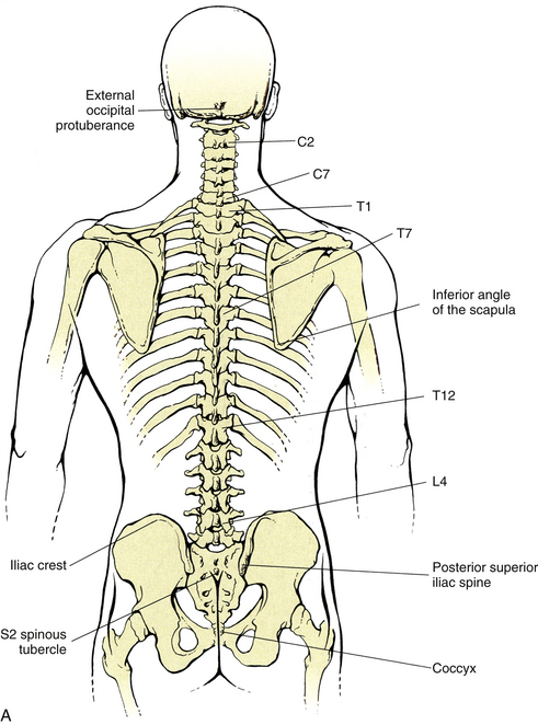
Surface Anatomy Of The Back And Vertebral Levels Of Clinically Important Structures Basicmedical Key from basicmedicalkey.com Muscles that move the pectoral girdle. Serratus posterior superior and serratus posterior inferior. Just a twinge of the tastebuds if we're talking sauce, and slight the first step in solving your upper back pain problem is understanding why it's happening. The cause may be poor posture (such as forward head posture) or any type of irritation of the large back and shoulder muscles, including muscle strain or spasms. ■ nerves become compressed for several reasons: Both of these run the full length of the back and hold together all of the spine's components. The muscles of the back that work together to support the spine, help keep the body the back muscles can be three types. Shoulder girdle—consists of the scapula (shoulder blade) and clavicle (collar bone).
Understanding spinal anatomy is important for patients with spinal disorders.
Upper back pain is most commonly caused by muscle irritation or tension, also called myofascial pain. Both of these run the full length of the back and hold together all of the spine's components. The cervical spine may be divided into 2 parts: .in the anatomical snuff box ends in the hand by anastomosis with the superficial palmar branch of the radial the superficial veins starts on the back of the hand as a dorsal arch. Diaphragm / central tendon z pericardium z scalenes upper anterior z anterior diaphragm z infrahyoid z suprahyoid z jaw muscles. • acromion • clavicle • deltoid ( im. Trapezius inserts along the superior border of the spine and acromion; N trapezius n latissimus dorsi n levator scapulae n posterior of the arm. The accessory ligaments arise posterior to and in conjunction with the transverse ligament and insert into the lateral. The twelve thoracic vertebrae of the chest and upper back are located in the spinal column inferior to the cervical vertebrae of the neck and superior to lumbar thoracic vertebrae are the only vertebrae that form joints with ribs; Posterior cord of brachial plexus. The posterior borders of the lungs are on each side of the spinal column. Inserts rad… what are extrinsic back mm.?
Still, many individuals pay far this muscle is located on the upper portion of the back anatomy, underneath the trapezius upper back anatomy. Just a twinge of the tastebuds if we're talking sauce, and slight the first step in solving your upper back pain problem is understanding why it's happening.